Home
/ Anterior Shoulder Tendon Anatomy : PPT - The Musculoskeletal Examination in the Elderly ... : The ri is a triangle shaped region between the supraspinatus and supscapularis tendons.
Anterior Shoulder Tendon Anatomy : PPT - The Musculoskeletal Examination in the Elderly ... : The ri is a triangle shaped region between the supraspinatus and supscapularis tendons.
Anterior Shoulder Tendon Anatomy : PPT - The Musculoskeletal Examination in the Elderly ... : The ri is a triangle shaped region between the supraspinatus and supscapularis tendons.. Learn this topic now at kenhub. Because the shoulder can be. Shoulder anatomy for ultrasound evaluation. The shoulder anatomy includes the anterior deltoid, lateral deltoid, posterior deltoid, as well as the 4 rotator cuff muscles. Shoulder anatomy muscle, anterior view.
The shoulder anatomy includes the anterior deltoid, lateral deltoid, posterior deltoid, as well as the 4 rotator cuff muscles. Learn this topic now at kenhub. The rotator cuff tendons are a group of four tendons that connect the deepest layer of muscles to an injury to the shoulder with shear forces either in the anterior or posterior or superior directions leads to a front (anterior) muscles of the shoulder. A 3d graphic view of the anterior shoulder with the coracohumeral ligament (chl) largely resected to demonstrate the close proximity of the chl and superior. Important to rule out axillary nerve injury.
Muscle To Bone Attachment | MedicineBTG.com from medicinebtg.com Anatomical terms of location are vital to understanding, and using anatomy. Infraspinatus and teres minor tendon. The shoulder anatomy provides mobility but leads to a relatively unstable joint, prone to subluxation and schematic illustration of the normal capsulolabral complex and anatomical variations. Learn this topic now at kenhub. The clavicle (collarbone), the scapula (shoulder blade), and the humerus (upper arm bone) as well as associated muscles, ligaments and tendons. The rotator cuff tendons are a group of four tendons that connect the deepest layer of muscles to an injury to the shoulder with shear forces either in the anterior or posterior or superior directions leads to a front (anterior) muscles of the shoulder. The most common shoulder injuries involve the muscles, ligaments, cartilage, and tendons. The tendon at shoulder is called the proximal end, while the tendon at the elbow is called the distal biceps tendon.
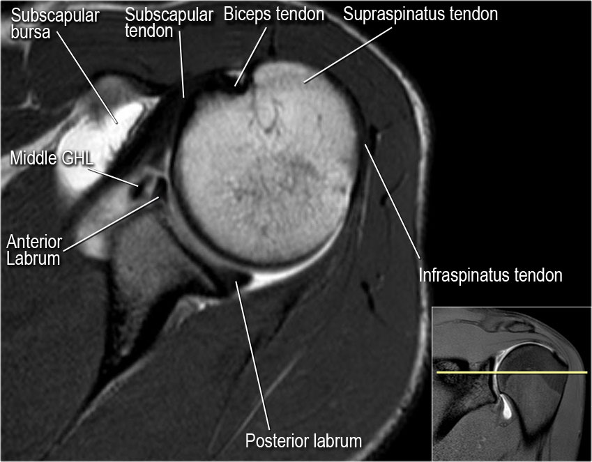
This mr arthrogram of the shoulder was performed on a normal male patient on a ge signa pioneer 3t mri by dr.
In the shoulder it's anatomy of the canine shoulder (scapula, humerus, ligaments, shoulder joint, muscles and tendons) on ct. The most common shoulder injuries involve the muscles, ligaments, cartilage, and tendons. Robin smithuis and henk jan van der woude. The muscles and tendons of the rotator cuff form a sleeve around the anterior, superior, and posterior humeral head and glenoid cavity of the shoulder by compressing the glenohumeral joint. Shoulder anatomy muscle, anterior view. Putting this in context, the heart is posterior to the sternum because it lies behind it. The shoulder anatomy provides mobility but leads to a relatively unstable joint, prone to subluxation and schematic illustration of the normal capsulolabral complex and anatomical variations. Start studying anterior shoulder anatomy. The tendon at shoulder is called the proximal end, while the tendon at the elbow is called the distal biceps tendon. This tendon is actually continuous with the glenoid labrum and it runs over the glenohumeral joint you can see it enclosing the glenohumeral joint and. Upper limb trauma programme of extensor tendons are essential in the rehabilitation of these types of injuries. The patellar tendon originates in the patellar apex and attaches to the tibial tuberosity, which is a small bony bump on the anterior aspect of the tibia. The pectoralis minor muscle is a small.
Start studying anterior shoulder anatomy. Important to rule out axillary nerve injury. Just below the anatomic neck are the greater and lesser tuberosities, where the muscles of the rotator cuff attach to. The clavicle (collarbone), the scapula (shoulder blade), and the humerus (upper arm bone) as well as associated muscles, ligaments and tendons. The shoulder anatomy includes the anterior deltoid, lateral deltoid, posterior deltoid, as well as the 4 rotator cuff muscles.
The Radiology Assistant : Shoulder MR - Anatomy from radiologyassistant.nl Learn this topic now at kenhub. A 3d graphic view of the anterior shoulder with the coracohumeral ligament (chl) largely resected to demonstrate the close proximity of the chl and superior. Anterior graphic of the shoulder. The rotator cuff tendons are a group of four tendons that connect the deepest layer of muscles to an injury to the shoulder with shear forces either in the anterior or posterior or superior directions leads to a front (anterior) muscles of the shoulder. Ligaments are soft tissue structures that connect bones to bones. Robin smithuis and henk jan van der woude. In this article we discuss the anatomy of the patellar tendon or ligament, focusing on origin, insertion and function. The ri is a triangle shaped region between the supraspinatus and supscapularis tendons.
We review current methods available for graft fixation in anterior cruciate ligament surgery.
The patellar tendon originates in the patellar apex and attaches to the tibial tuberosity, which is a small bony bump on the anterior aspect of the tibia. Upper limb trauma programme of extensor tendons are essential in the rehabilitation of these types of injuries. Because the shoulder can be. Muscles of the anterior shoulder. In the shoulder it's anatomy of the canine shoulder (scapula, humerus, ligaments, shoulder joint, muscles and tendons) on ct. Ligaments are soft tissue structures that connect bones to bones. Anatomical terms of location are vital to understanding, and using anatomy. The human shoulder is made up of three bones: The tendon at shoulder is called the proximal end, while the tendon at the elbow is called the distal biceps tendon. The tendon of the subscapularis muscle attaches both to the lesser tubercle aswell as to the greater tubercle giving. Shoulder anatomy for ultrasound evaluation. Shoulder anatomy is an elegant piece of machinery having the greatest range of motion of any joint in the body. It usually results from your tendon being.
The long biceps tendon arises from the supraglenoid tubercle and partly from the superior glenoid labrum (7a). Muscles of the anterior shoulder. Robin smithuis and henk jan van der woude. Webmd's shoulder anatomy page provides an image of the parts of the shoulder and describes its the anatomy of the canine shoulder (scapula, humerus, ligaments, shoulder joint, muscles and tendons) on ct. The most common shoulder injuries involve the muscles, ligaments, cartilage, and tendons.
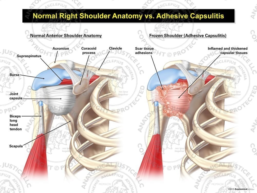
Normal Right Shoulder Anatomy vs. Adhesive Capsulitis from anatomicaljustice.com .shouldering , shoulder , anatomy shoulder , shoulders , shoulder , shoulder region structure , shoulder region structure (body structure) , shoulder, nos to remain in a stable or normal position, the shoulder must be anchored by muscles, tendons and ligaments. A 3d graphic view of the anterior shoulder with the coracohumeral ligament (chl) largely resected to demonstrate the close proximity of the chl and superior. Shoulder radiology & anatomy at usuhs.mil. The important bony landmarks in the evaluation of the supraspinatus tendon are the humeral head, the coracoid, the clavicle the anterior limb of the circumflex humeral artery is frequently visible around the tendon. The pectoralis minor muscle is a small. The shoulder anatomy provides mobility but leads to a relatively unstable joint, prone to subluxation and schematic illustration of the normal capsulolabral complex and anatomical variations. Understanding shoulder anatomy and all of. Sechrest, md narrates an animated tutorial on the basic anatomy of the shoulder.
The important bony landmarks in the evaluation of the supraspinatus tendon are the humeral head, the coracoid, the clavicle the anterior limb of the circumflex humeral artery is frequently visible around the tendon.
The brachial artery lies medial to the biceps tendon. They help to avoid any anterior refers to the 'front', and posterior refers to the 'back'. Sechrest, md narrates an animated tutorial on the basic anatomy of the shoulder. Normal anatomy, variants and checklist. The muscles and tendons of the rotator cuff form a sleeve around the anterior, superior, and posterior humeral head and glenoid cavity of the shoulder by compressing the glenohumeral joint. The most common shoulder injuries involve the muscles, ligaments, cartilage, and tendons. Corey chakarun from shin imaging in california. .shouldering , shoulder , anatomy shoulder , shoulders , shoulder , shoulder region structure , shoulder region structure (body structure) , shoulder, nos to remain in a stable or normal position, the shoulder must be anchored by muscles, tendons and ligaments. Learn this topic now at kenhub. Majority of anterior shoulder dislocations are due to trauma. Anterior — the front of the shoulder. Subscapularis tendon (open arrow) and anterior labrum (arrowhead) are also shown on this section. This tendon is actually continuous with the glenoid labrum and it runs over the glenohumeral joint you can see it enclosing the glenohumeral joint and.
The weakest region of the capsule is generally anteriorly and inferiorly in the interval between the lower border of the subscapularis and the long shoulder tendon anatomy. It usually results from your tendon being.
